
Guided Dental Implant Placement & Immediate Restoration | Clinical Case Study
Case Study: Guided Immediate Implant Placement and Anterior Rehabilitation using Warau Digital Workflow.
At a Glance: Clinical Case Summary
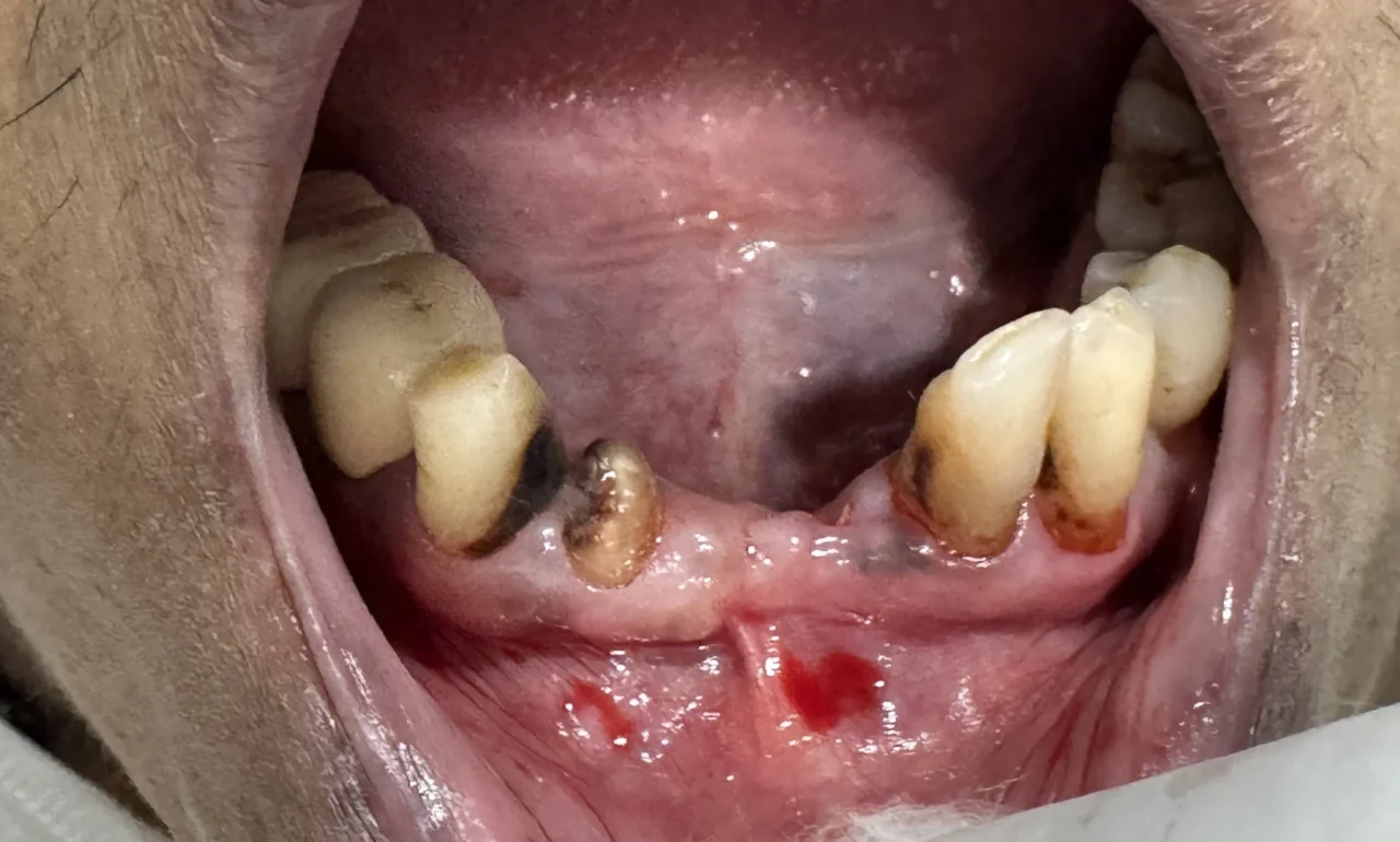
- Patient Presentation: Non-restorable failing mandibular anterior (lower front) teeth with localized bone loss, impaired function, and aesthetic concerns.
- Diagnosis: Compromised lower anterior teeth requiring extraction and immediate fixed prosthetic rehabilitation.
- Procedure:
- 3D CBCT and intraoral digital scan for digital 3D implant planning
- Atraumatic extraction of failing anterior roots
- Computer-guided immediate dental implant placement using a custom 3D-printed surgical template
- Immediate temporary screw retained prosthesis.
- Prosthesis: Fixed multi-unit implant-supported bridge custom-fabricated on printed master models.
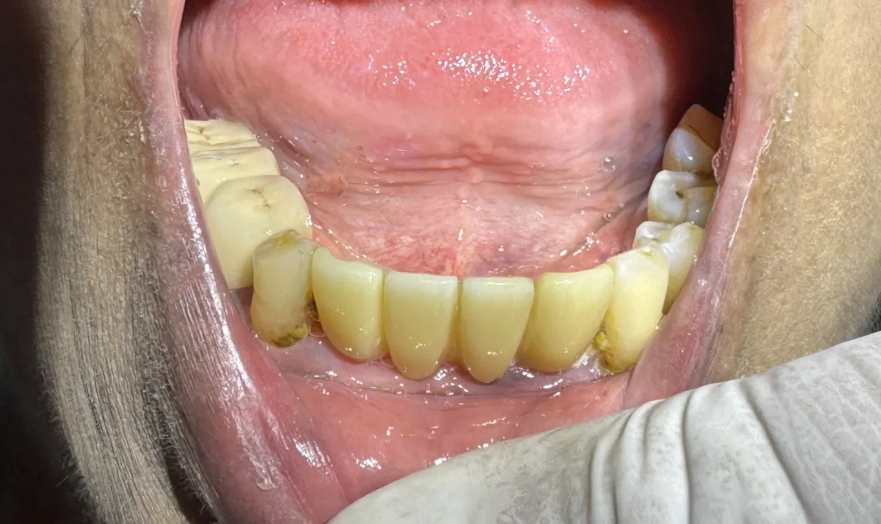
- Primary Outcome: Complete restoration of natural aesthetics, optimal soft tissue adaptation, balanced bite occlusion, and high patient satisfaction.
- Key Advantage: Guided surgery using Warau Digital workflow ensured precise 3D implant positioning, reduced treatment time, and preserved natural bone and gum contours.
1. Introduction & Patient Presentation
Missing or non-restorable mandibular anterior teeth pose both functional and aesthetic challenges. A patient presented to our dental clinic presenting with failing lower anterior dentition, localized mobility, and aesthetic distress. The primary clinical goal was to restore speech, chewing efficiency, and lower-third facial aesthetics using a digitally planned, guided surgical approach for dental implant rehabilitation.
2. Step 1: Comprehensive Data Collection & Diagnostics
A predictable implant treatment starts with meticulous diagnostic data collection:
- Clinical Examination: Detailed evaluation of oral hygiene, periodontal health, smile line, gingival biotype, interarch space, and occlusal scheme.
- 3D CBCT Imaging (Cone Beam Computed Tomography): High-resolution CBCT scanning to assess bone height, bone width, bone quality/density, and proximity to anatomical structures (such as the mental foramen and lingual cortical plate).
- Intraoral Scanning (IOS) / Digital Impressions: High-precision optical impressions taken of the maxillary and mandibular arches to capture soft tissue contours, tooth morphology, and existing occlusion.
- Extraoral & Intraoral Photography: Pre-operative photographic baseline taken for aesthetic analysis and digital smile design integration.
3. Step 2: Virtual 3D Planning & Surgical Guide Design
Combining the CBCT data (DICOM format) with the optical intraoral scan (STL format) allowed for comprehensive virtual surgical simulation:
- DICOM-STL Merging: Precise registration of hard tissue (CBCT) and soft tissue/teeth contours (IOS) in computer-aided implant planning software.
- Virtual Implant Selection & Positioning: Prosthetically driven placement of virtual implants, ensuring optimal 3D axis orientation for screw vs. cement retention, ideal crestal bone depth, and safe distance from adjacent roots.
- 3D Printed Surgical Guide Fabrication: Designing a tooth-supported surgical template for precise depth and angular control during drilling, followed by high-precision 3D printing and biocompatible post-processing.
4. Step 3: Surgical Execution (Atraumatic Extraction & Guided Drilling)
- Atraumatic Tooth Extraction: Careful extraction of non-restorable lower front teeth using periotomes to completely preserve surrounding buccal and lingual alveolar bone plates.
- Socket Debridement: Thorough curettage and copious saline irrigation of fresh extraction sockets to remove granulation tissue.
- Surgical Guide Placement: Insertion and fit verification of the custom 3D-printed surgical template onto adjacent anchor teeth to guarantee absolute stability.
- Guided Osteotomy & Implant Insertion: Sequenced osteotomy preparation using the guided drill kit, followed by insertion of dental implants into pre-planned positions with high initial torque (primary stability).
- Position Verification: Depth gauges and direction pegs utilized to cross-verify structural alignment and inter-implant parallelism prior to final seating.
5. Step 4: Laboratory Fabrication & Provisionalization
- Impression / Scan Post-Placement: Post-surgical digital scan or micro-impression taken to capture exact 3D implant positions relative to soft tissues.
- Provisional Rehabilitation: Placement of a protective temporary restoration or bridge during the initial osseointegration and soft-tissue healing period.
- Master Cast & Model Printing: Fabrication of high-precision 3D resin master models incorporating implant analogs for custom laboratory processing.
- Prosthetic Finishing: CAD/CAM fabrication of a multi-unit ceramic-fused-to-metal or monolithic zirconia bridge with individualized gingival contouring and natural enamel translucency.
6. Step 5: Final Delivery & Occlusal Adjustment
- Try-In & Passivity Check: Seat verification of the custom multi-unit prosthesis to ensure absolute passive fit over implant abutments without tension.
- Torque Protocol & Seating: Final tightening of prosthetic screws or cementation according to manufacturer-recommended torque guidelines.
- Occlusal Calibration: Verification of balanced occlusion, eliminating premature contacts during protrusive and lateral excursions.
- Post-Operative Outcome: Seamless integration with natural teeth, excellent tissue adaptation, complete restoration of masticatory function, and superior aesthetic results.
Key Benefits for Patients
- Computer-Guided Safety: 3D digital planning eliminates guesswork, protecting vital structures and ensuring exact implant positioning.
- Preserved Facial Structure: Immediate post-extraction implant placement maintains natural jawbone density and gum line contours.
- Faster Recovery: Guided minimally invasive procedures typically lead to less swelling, reduced pain, and faster healing times.
- Long-Lasting Aesthetic Comfort: Custom-designed prosthetic restorations feel, look, and function just like natural teeth.